Breast Lump Treatment - Your One Stop Centre
We will relief you from your worries
From blood works, ultrasound scan, tissue biopsy to breast lump excision surgery
Common Causes of Breast Lump and Swelling
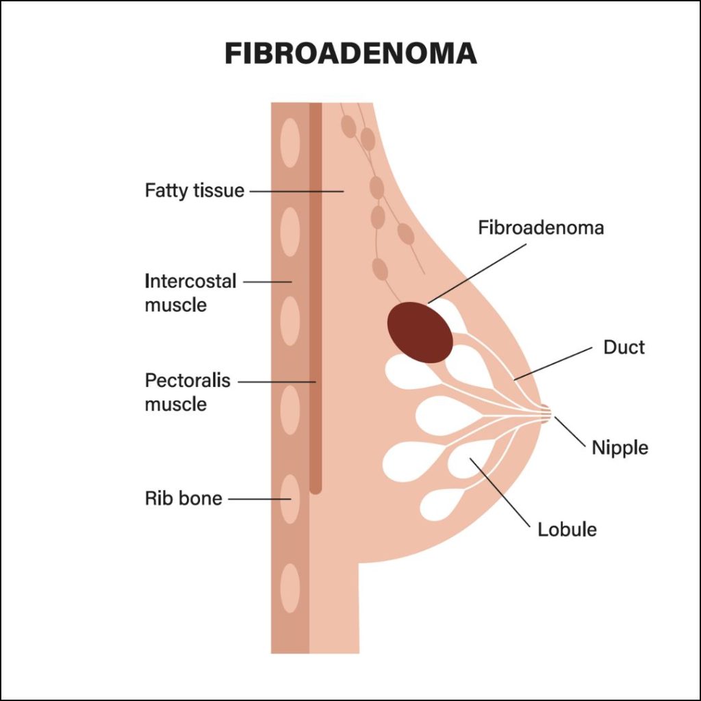
1. Fibroadenoma
- Fibroadenoma is a painless, one-sided, non-cancerous breast lump that usually occurs between the age of 15 to 35 years old.
- Usually small (1-2cm), has a smooth surface and very mobile (often describe as mouse under the skin)
- Can grow during pregnancy and shrink after menopause.
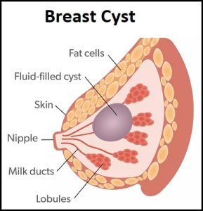
2. Breast cyst
- More common than fibroadenomas. Usually in the age group between 30 to 50 years old.
- Occurs due to involution or shrinking of breast tissue and the milk gland turning into cyst.
- It is a benign condition (non-cancerous). Most breast cyst are asymptomatic (no symptoms) and are usually detected during routine breast examination.
- Some larger breast cyst can present as pain, lump or nipple discharge.
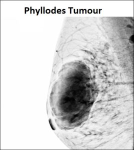
3. Phyllodes tumour
- Rare form of breast lump. It usually occurs in middle age women between 40 to 50 years old.
- It usually present as a firm, rapidly enlarging breast lump that stretch the overlying skin and associated with distension of the nearby veins.
- It usually involves only 1 side of the breast.
- Can vary in size from 1cm to as big as 40cm and can occupy the entire breast.
- It is a benign condition but has the potential to turn into malignant (cancerous) tumour.
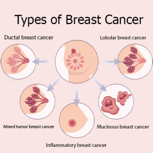
4. Breast cancer
- Breast cancer is the most commonly diagnosed cancer in female population.
- It usually present as a painless lump and can be associated with
- Bleeding or unusual nipple discharge
- Skin changes over the lump such as dimpling, puckering.
- The lump has an irregular border and fixed to the adjacent tissue.
- Retracted or pulled in nipple.
- Thickening, ulcerated or rash over the skin.
- The risk factors for breast cancers are:
- Age above 40 years old.
- Family history of breast cancer.
- Early menstruation or late menopause.
- Late childbirth.

I have a breast lump. What should I do next?
The first thing that you should do is not to panic. Most breast lump are benign in nature. Arrange for an appointment with our female doctor to have a further look into the breast lump
1. Consultation and physical examination
Although seems trivial, an appointment with doctor can reveal a lot of information during the consultation and examination session. Our doctor will ask further information such as associated symptoms, family history and any previous investigation done. 80% of diagnosis of a breast lump can be derived during the consultation and physical examination session.
2. Imaging study (Ultrasound Scan)
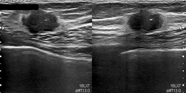
Sometimes, more information is needed before doctor can conclude what is the diagnosis. Further imaging study will help to differentiate a benign breast lump with a malignant growth. Here at Klinik Safa, we will do ultrasound scan of the lump to look for sign of malignancy, such as having a mixed echogenicity, irregular border of a lump and calcification within the lump.
3. Tissue biopsy
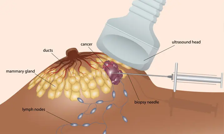
If all of the above still cannot determine whether a lump is benign or malignant, the gold standard is by Histopathological Examination or known as HPE, where a sample of the tissue will be sent to the lab to be viewed under the microscope. This can be done using a small biopsy needle. The procedure is done under local anaesthesia so there is no pain. The result will be out within 3-5 working days and our doctor will explain further on the next course of action.
Treatment Options
1. Conservative or Watchful waiting
Most benign breast lumps do not require any surgical intervention. Periodic ultrasound scan and blood tumour marker monitoring will be done to ensure that the lesion is not dangerous.
2. Excision biopsy
Benign breast lump that causes pain or for cosmetic reason can be removed under local anaesthesia. The procedure can be done in the clinic and does not require hospital admission. The removed tissue will be examined after the removal. If it shows signs of malignancy, it will be sent to the lab for Histopathological Examination (HPE) to confirm the type of tissue and whether it is malignant or not.
2. Mastectomy
Mastectomy is the removal of the whole breast tissue. This is usually done for malignant breast cancer to ensure that no cancer cell are left behind. It is done under general anaesthesia where you will be put to sleep during the procedure. It requires hospital admssion and cannot be done in the clinic or daycare setting.
